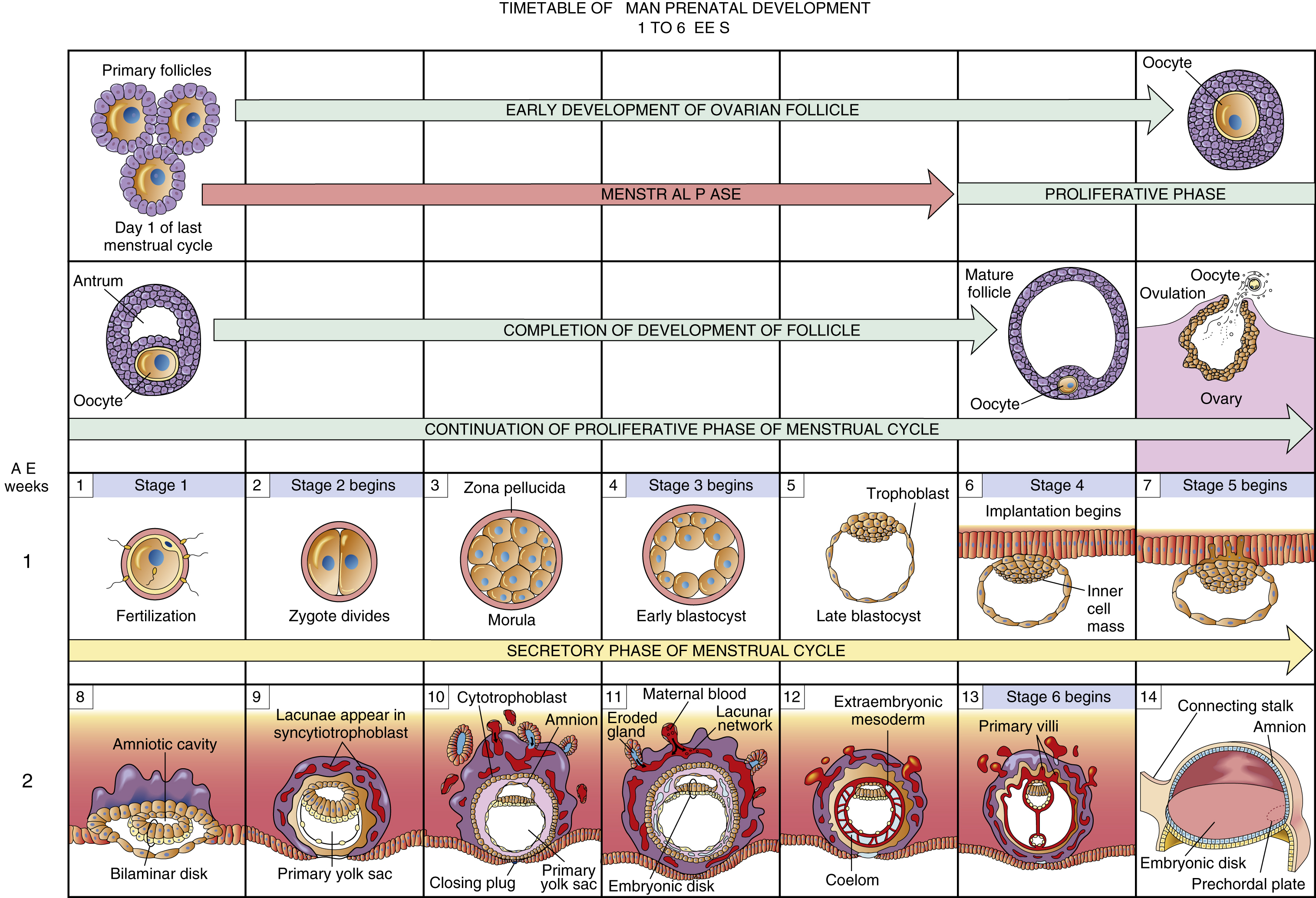
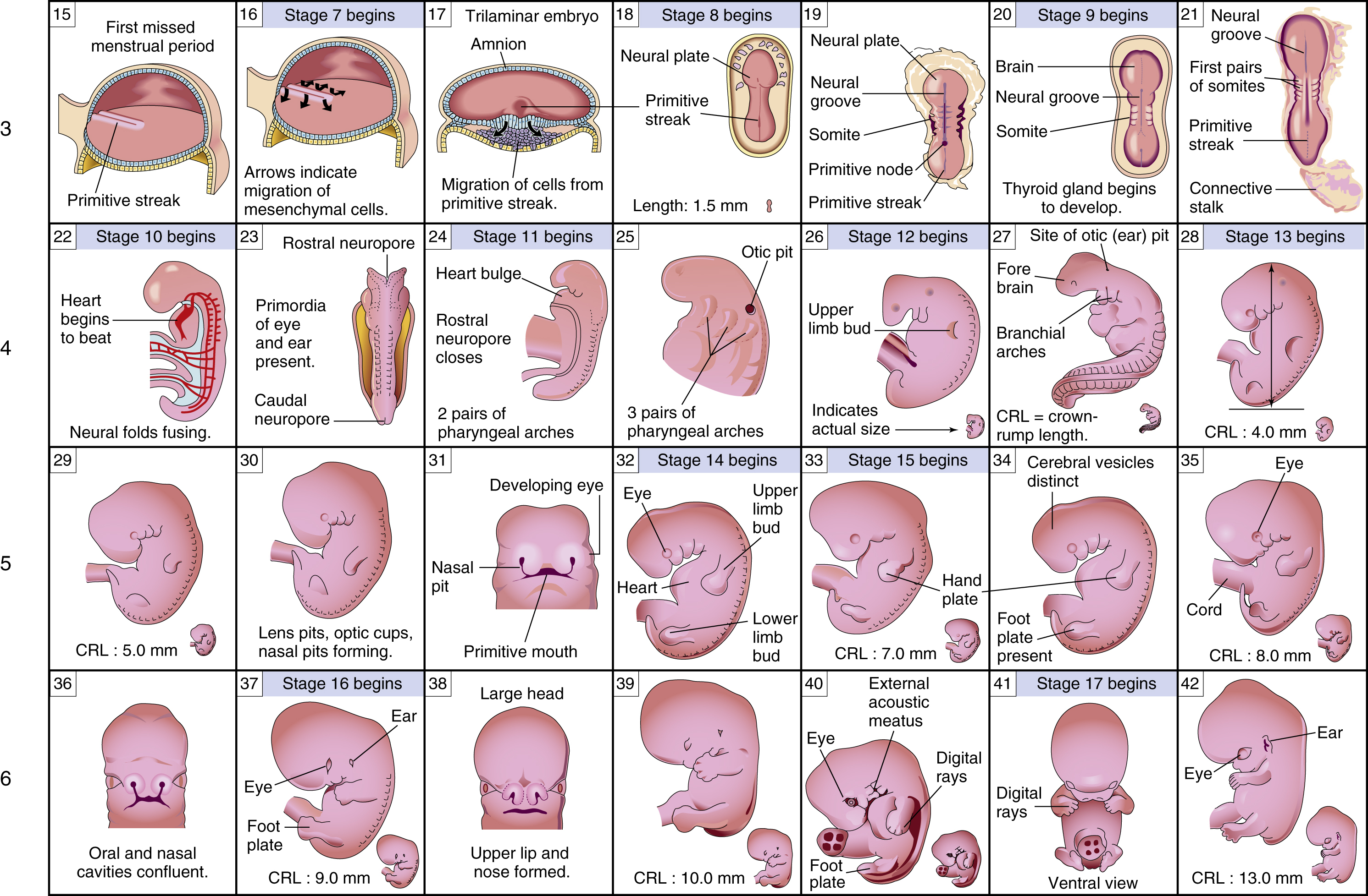
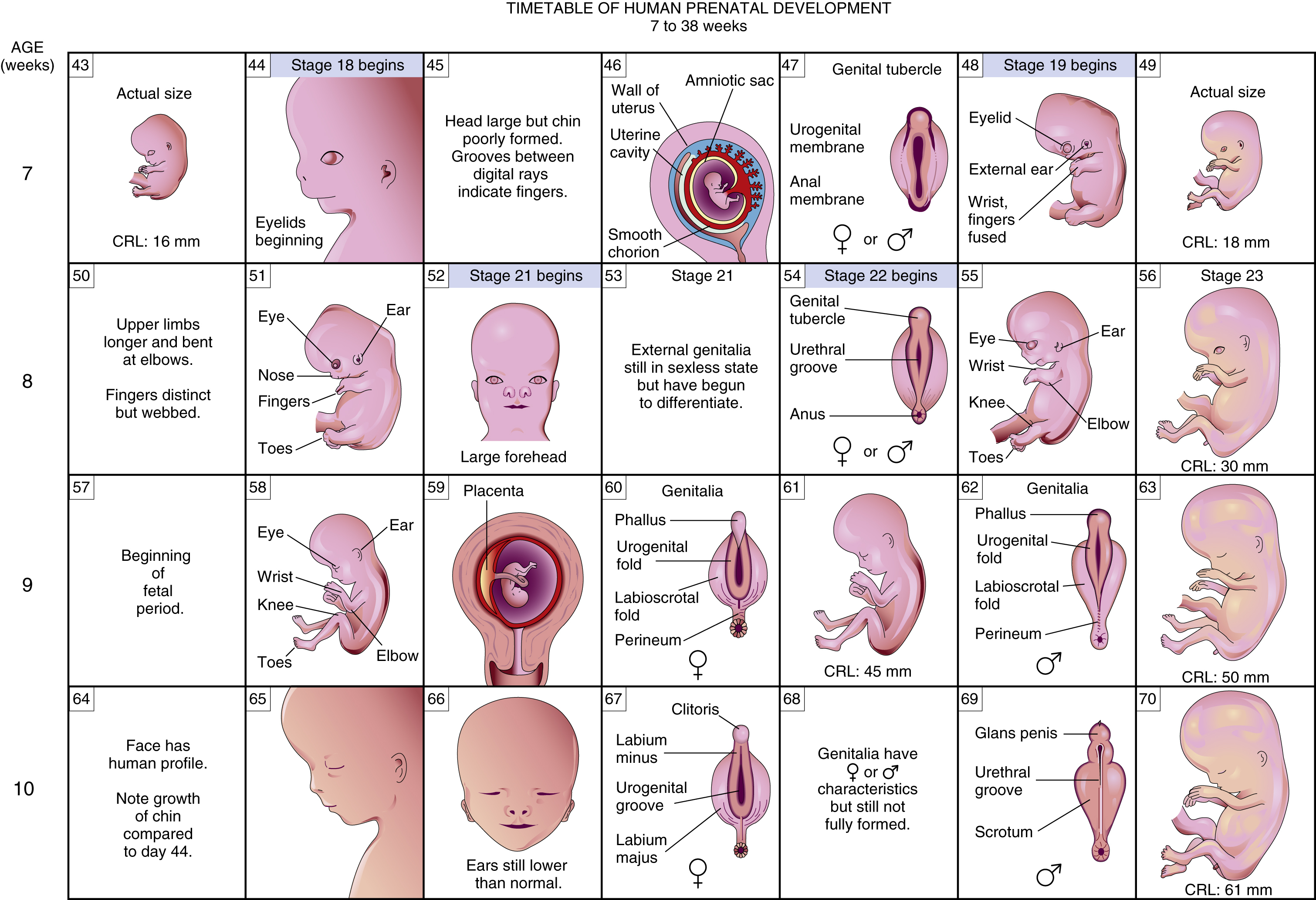
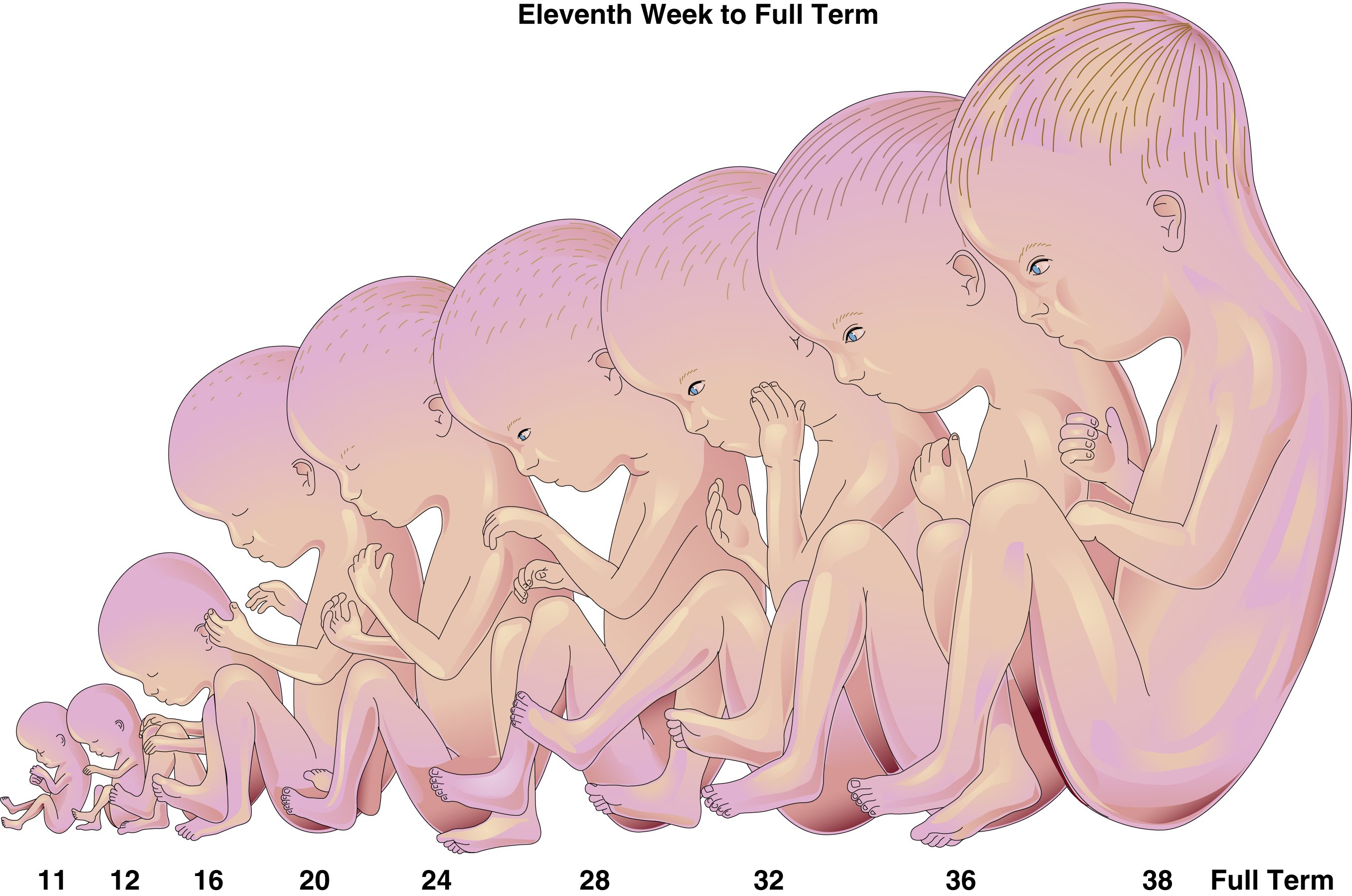
prenatal development, the entire process of growth, maturation, differentiation, and development that occurs between conception and birth. On approximately the 14th day before the next expected menstrual period ovulation usually occurs. If the egg is fertilized, it immediately begins the course to fetal maturity and birth. During the first 14 days the fertilized ovum undergoes cell division several times, becoming a morula and then a blastocyst that is able to implant in the uterine wall. From the beginning of the third to the end of the seventh week of embryonic development, implantation deepens and completes. Primitive uteroplacental circulation originates between the enlarging trophoblast and the maternal endometrial tissue of the uterus. The amniotic cavity appears as an opening between the inner cell mass and the invading trophoblast. A thin lining in the cavity becomes the amnion. At this point the embryo is a two-layered embryonic disk composed of an ectoderm and an endoderm. As the disk thickens in the middle, giving rise to the third cell layer, or mesoderm, the basic structural systems of the body begin to form. The neural tube develops as a precursor of the central nervous system in the midline of the cranial part of the ectoderm. Primitive blood vessels and blood cells, a heart tube, and umbilical vessels are formed and begin to function. Arm and leg buds may appear, and rudimentary gut, lungs, and kidneys form. By the fifth week the brain has begun to grow rapidly, the heart tube is divided into chambers, the palate and the upper lip are forming, and the urogenital system is developing. By the end of the seventh week all essential systems are present. The period from the eighth week to birth is called the fetal stage. From the 8th to the 10th week the fetus continues to grow and development is rapid. The head is almost half of its total length, and arms, legs, and face are clearly recognizable. The fetus floats in the amniotic fluid of the amniotic sac within the uterus; the umbilical vessels in the cord extend to a rapidly growing placenta. By the twelfth week the facial features are formed and the eyelids are present but not yet closed because they have not divided into upper and lower eyelids. The palate is fusing, a neck connects the large head and the body, and tooth buds and nailbeds have begun to form. Identification of the external genitalia is possible for the first time. From the13th to the 16th week the arms, legs, and trunk grow rapidly, and the fetus is active. Scalp hair develops. The skeleton of the fetus is calcified and may be seen on an x-ray film. A sonogram sometimes detects respiratory movements. Between the 17th and the 20th week of pregnancy the mother usually first feels the baby move. The fetus looks like a very small baby at this time. There are eyebrows and tiny nipples; during fetoscopic examination the fetus has been seen and photographed sucking its thumb and grasping its own umbilical cord. At the 24th week the external ears are smooth and soft and the skin is wrinkled and translucent. The body is covered with lanugo and vernix and weighs a little more than 1 pound. At 28 weeks subcutaneous fat begins to develop, fingernails and toenails are present, the eyelids are separate, the eyes may open, scalp hair is well developed, and in males the testes are at the internal inguinal ring or below. In a modern neonatal intensive care unit most of the babies born at 28 weeks survive. By the 32nd week the fetus weighs between 3 and 4 pounds. The hair is fine and woolly, the fingernails and toenails have grown to the tips of the fingers and toes, and there are one or two creases on the anterior part of the soles of the feet. The areolae of the breasts are visible but flat. In females the clitoris is prominent and the labia majora are small and separated. At 36 weeks the body and the limbs are fuller and more rounded, creases involve the anterior two thirds of the soles, and the skin is thicker and less translucent. As the fetus reaches term, between 38 and 42 weeks, the vernix decreases, and the ear cartilage is developed. In males the testes are in the scrotum. In females the labia majora meet in the midline and cover the labia minora and the clitoris. At 40 weeks the average fetus weighs 7 ¼ pounds and is between 19 and 22 inches long. Prenatal development may be adversely affected by several factors. Between 2 and 14 weeks of gestation, ionizing radiation and some drugs may have profound effects on morphological and functional development. During the first 10 days of development, any damage usually kills the conceptus. Various viruses, malnutrition, trauma, or maternal disease may also affect the morphological development of a rapidly differentiating structure or organ during the embryological or early fetal stage. After 14 weeks, when all of the organs, systems, and body parts have formed, any adverse effects are largely functional; major morphological damage does not occur.


