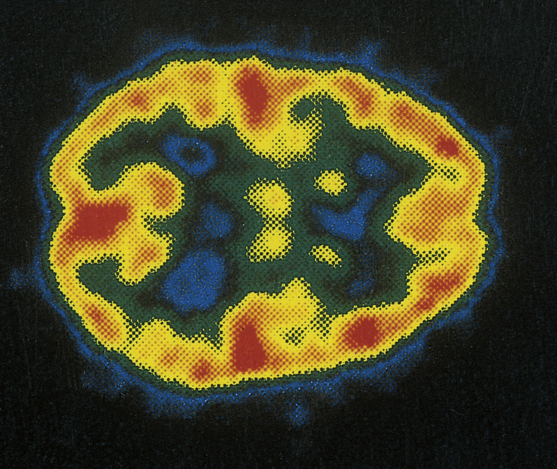
positron emission tomography (PET) [L, positivus + Gk, elektron, amber; L, emittere, to send out; Gk, tome, section, graphein, to record] , a computerized radiographic technique that uses radioactive substances to examine the metabolic activity of various body structures. The patient either inhales or is injected with a metabolically important substance such as glucose, carrying a radioactive element that emits positively charged particles, or positrons. When the positrons combine with electrons normally found in the cells of the body, gamma rays are emitted. The electronic circuitry and computers of the PET device detect the gamma rays and construct color-coded images that indicate the intensity of metabolic activity throughout the organ involved. The radioactive isotopes used in PET are very short-lived, so that patients undergoing a PET scan are exposed to very small amounts of radiation. Researchers use PET to examine blood flow and the metabolism of the heart and blood vessels, to study and diagnose cancer, and to investigate the biochemical activity of the brain.

