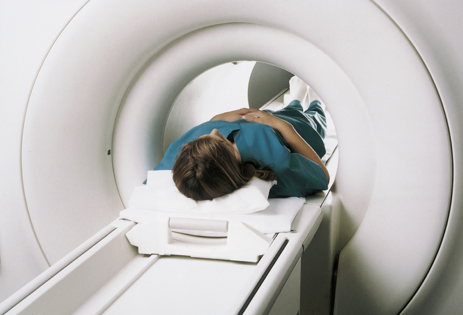
magnetic resonance imaging (MRI) [Gk, magnesia, lodestone, resonare, to sound again, imago, image] , medical imaging based on the resonance of atomic nuclei in a strong magnetic field. The field causes those nuclei with an odd number of protons to align and rotate around the axis of the field. Application of a radiofrequency pulse causes the protons to resonate. When the pulse is terminated, the protons “relax” back toward equilibrium. As they do so, they release energy that is detected as a radio signal. Analysis of the amplitude and frequency of the signal yields information about the number and position of nuclei in the tissue, from which the image is produced. MRI is the method of choice for a growing number of disease processes. Among its advantages are its superior soft-tissue contrast resolution, ability to image in multiple planes, and lack of ionizing radiation hazards. MRI is regarded as superior to computed tomography for most central nervous system abnormalities, particularly those of the posterior fossa, brainstem, and spinal cord. It has also become an important tool in musculoskeletal and pelvic imaging. The procedure usually does not require a contrast medium but may use an IV injection of gadolinium. About 15% of patients require an anxiolytic to overcome claustrophobia during MRI, and children may need a sedative as well. Patients must remain motionless for high-quality imaging. Images also may be degraded by motions related to heart contractions, respiration, and bowel peristalsis. Contraindications to MRI are pacemakers, metallic aneurysm clips, and some metallic prostheses and foreign objects. Compare open magnetic resonance imaging. Formerly called zeugmatography. See also magnetic resonance.

