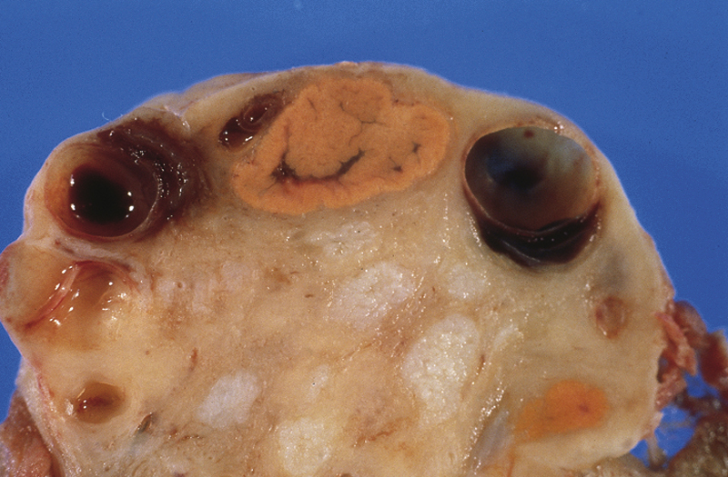
corpus luteum /kôr″pəs lo̅o̅″tē·əm/ pl. corpora lutea [L, corpus, body, luteus, yellow] , an anatomical structure on the ovary’s surface, consisting of a spheroid of yellowish tissue 1 to 2 cm in diameter that grows within the ruptured ovarian follicle after ovulation. The pleated wall of the collapsed follicle is made up of several layers of granulosa cells that grow toward the center of the cavity to form the structure. During a woman’s reproductive years, a corpus luteum forms after every ovulation. It acts as a short-lived endocrine organ that secretes progesterone, which serves to maintain the decidual layer of the uterine endometrium in the richly vascular state necessary for implantation and pregnancy. If conception occurs, the corpus luteum grows and secretes increasing amounts of progesterone. It reaches its maximum function and size (2 to 3 cm) at 10 to 12 weeks of gestation. It persists, slowly diminishing in size and function, until 6 months after the onset of gestation. During the 2 weeks before menstruation, the corpus luteum secretes progesterone in decreasing amounts, atrophies, undergoes fibrotic degeneration, and becomes a pale spot on the surface of the ovary. Compare corpus albicans.

