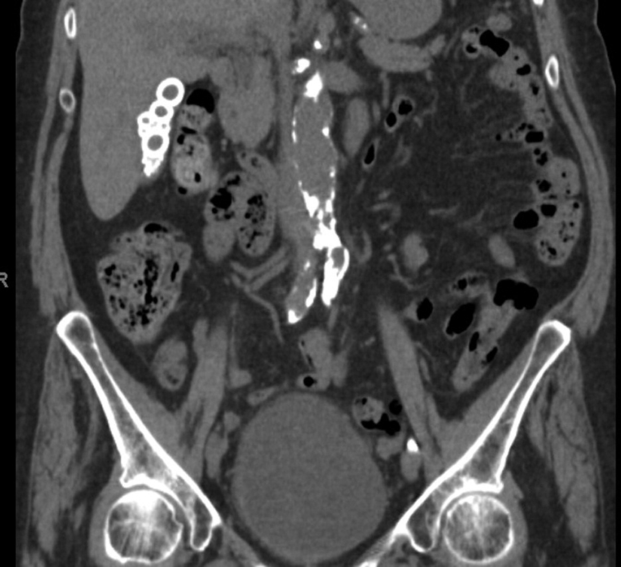
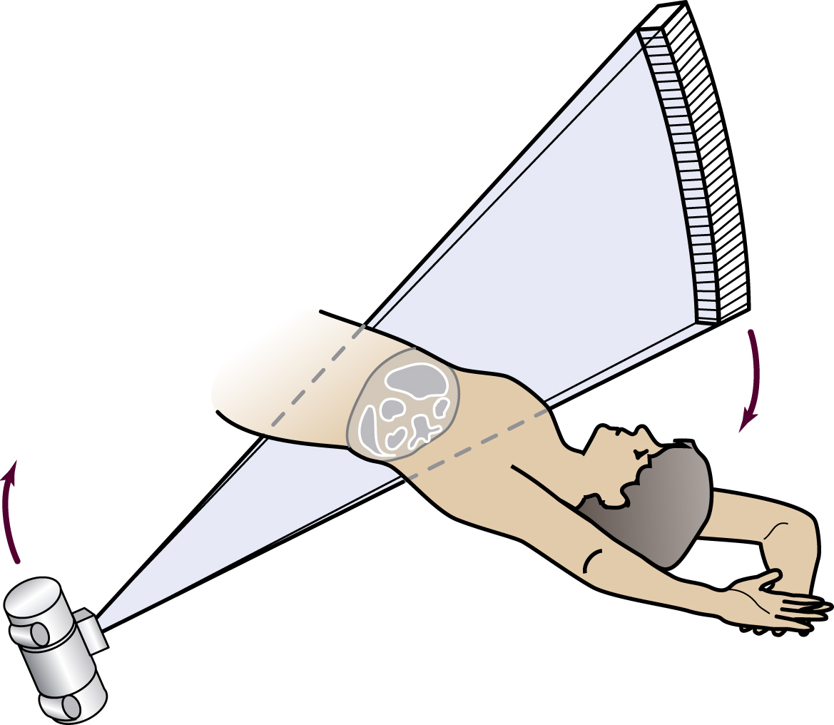
computed tomography (CT) /kəmpyo̅o̅″tid/ , 1. a radiographic technique that produces an image of a detailed cross-section of tissue. The procedure, first used in 1972, is painless and noninvasive and requires no special preparation. It is 100 times more sensitive than conventional radiography. CT uses a narrowly collimated beam of x-rays that rotates in a full arc around the patient to image the body in cross-sectional slices. An array of detectors, positioned at several angles, records those x-rays that pass through the body. The image is created by a computer that uses multiple attenuation readings taken around the periphery of the body part. The computer calculates tissue absorption and produces a representation of the tissues that demonstrates the densities of the various structures. Tumor masses, infractions, bone displacement, and accumulations of fluid may be detected. For cardiological examination, ultrafast CT is electrocardiogram-triggered and allows visualization of cardiac function and blood flow. Because modern CT equipment does not involve motion of the x-ray tube, heat loading is not a problem and multilevel images can be acquired in a very short time, sometimes during a single held breath. During a period of two held breaths, as many as 50 continuous tomographic images can be produced in a single-slice mode. Formerly called computerized axial tomography. 2. in dentistry, also called cone-beam imaging or cone-beam computed tomography (CBCT). Often used to view the temporomandibular joint area or a presurgical dental implant site.


