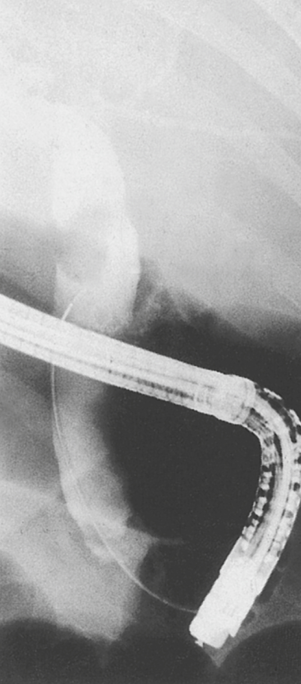
cholangiography /kōlan′jē·og″rəfē/ , a special roentgenographic test procedure for outlining the major bile ducts by the IV injection or direct instillation of a radiopaque contrast material. See also cholecystography. ▪ METHOD: For IV cholangiography the contrast agent is given slowly by vein, and x-ray films are taken of the region of the gallbladder. Operative and postoperative cholangiography use the injection of contrast material into the common bile duct via a drainage T-tube inserted during surgery to reveal any small, residual gallstones that are present. In percutaneous transhepatic cholangiography the contrast material is injected through a long needle or needle catheter, which is introduced directly through the skin into the substance of the liver. Endoscopic retrograde cholangiography is accomplished by cannulating the ampulla of Vater through a flexible fiberoptic duodenoscope and instilling radiopaque material directly into the common bile duct. ▪ INTERVENTIONS: IV cholangiography cannot be used in the presence of severe liver disease or jaundice because the dye will not be concentrated and excreted into the bile. The patient fasts, and fluids are restricted overnight. An early morning cleansing enema is given, usually followed by a sedative. The patient is warned about a brief burning sensation that occurs as the dye is injected. For percutaneous transhepatic cholangiography, sedative premedication is often ordered and a local anesthetic injected at the site of needle puncture. Appropriate evaluation for bleeding tendencies must be carried out before percutaneous transhepatic cholangiography. Bile peritonitis is occasionally a complication of T-tube or percutaneous cholangiography, and close nursing observation is essential after the test is completed. For endoscopic retrograde cholangiography, nothing is given by mouth after midnight, an explanation is given to the patient, dentures are removed, and, to permit administration of medications, IV infusion is begun. The endoscope is passed with the patient in the left lateral position; then the patient is turned to the prone position, the ampulla is cannulated, the dye is injected, and films are taken. Vital signs are observed and the patient is given a light meal 2 to 4 hours after the procedure. ▪ OUTCOME CRITERIA: The resulting cholangiograms from any of these procedures are examined for unobstructed outlining of the biliary system. Calculi may be noted as shadows within the opaque medium.

