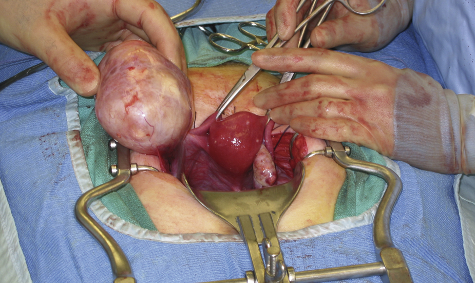
ovarian carcinoma, a malignant neoplasm of the ovaries rarely detected in the early stage and usually far advanced when diagnosed. It occurs frequently in the fifth decade of life. Ovarian cancer appears to be increasing in the United States. Risk factors of the disease are infertility, nulliparity or low parity, delayed childbearing, repeated spontaneous abortion, endometriosis, group A blood type, family history, previous history of breast or colorectal cancer, previous irradiation of pelvic organs, and exposure to chemical carcinogens such as asbestos and talc. Characteristics of the disease as it advances are abdominal swelling and discomfort, abnormal vaginal bleeding, weight loss, dysuria or abnormal frequency of urination, constipation, and a palpable ovarian mass, especially in postmenopausal women. Most ovarian carcinomas are papillary or serous, followed in frequency by mucinous, endometrial, and undifferentiated cancers. In many cases the cancer spreads over the surface of the peritoneum, and, early in the course of the lesion, tumor cells invade the lymphatic vessels under the diaphragm and the paraaortic nodes. Also called ovarian cancer. ▪ OBSERVATIONS: After an insidious onset and asymptomatic period, the tumor may become evident as a palpable abdominal or pelvic mass accompanied by irregular or excessive menses or postmenopausal bleeding. In advanced cases the patient may have ascites, edema of the legs, and pain in the abdomen and the backs of the legs. Papanicolaou smear may show malignant cells if the tumor is advanced; CA-125 may be elevated; an ultrasonic examination can demonstrate an ovarian mass but does not distinguish between a benign and malignant lesion. A computed tomographic scan may be useful in detecting ovarian cancer, but a definitive diagnosis requires surgical exploration. ▪ INTERVENTIONS: Approximately half of the tumors diagnosed are unresectable. Treatment of resectable lesions consists of total abdominal hysterectomy, removal of both ovaries and tubes, omentectomy, and biopsies of any suspicious sites, especially in the liver and diaphragm. Chemotherapeutic agents that may be administered after surgery include chlorambucil, cisplatin, cyclophosphamide, melphalan, taxol, docetaxel, and thiotepa. Radiation therapy may be used alone or in conjunction with chemotherapy. “Second-look” surgery is usually performed 1 year after chemotherapy to confirm the eradication of the tumor. ▪ PATIENT CARE CONSIDERATIONS: Regular yearly pelvic examinations after 40 years of age contribute significantly to early diagnosis and the possibility of curative treatment.

